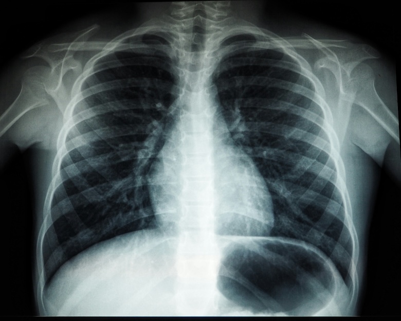
In the realm of cancer diagnosis, medical imaging is a pivotal tool. It offers in-depth visuals of the body’s interior, thereby facilitating early detection, staging, monitoring treatment progress, and evaluating recurrence. A variety of imaging tests are employed for diagnosing cancer:
- Computed Tomography (CT) Scan: This generates three-dimensional images of the body, enabling visualization of bones, organs, tumors, and soft tissues.
- Magnetic Resonance Imaging (MRI): Utilizing radio waves and a potent magnet, this technique produces detailed images showcasing structures such as tumors.
- Nuclear Medicine Scan: This method uses radioactive tracers to identify tumors and evaluate their metabolic activity.
- Positron Emission Tomography (PET) Scan: This reveals areas with heightened glucose uptake which often signifies the presence of cancer cells.
- Ultrasound: By employing sound waves, it forms images and evaluates tissue elasticity.
Imaging tests hold significant importance for early detection, staging, planning therapy strategies, assessing treatment responses and ongoing patient monitoring. They offer vital data that helps determine the location, size and spread of cancer thus influencing treatment choices and prognosis evaluation. The amalgamation of anatomical and functional imaging techniques like CT scans, MRIs, PET scans and SPECT provides an all-encompassing understanding of cancer at both structural and molecular levels.

